Key points on CAKUT syndrome
- CAKUT, or congenital anomalies of the kidney and urinary tract, refers to a range of structural birth defects in the kidneys, ureters, bladder, or urethra that affect about 1 in every 200 to 500 newborns, though exact numbers vary by population.
- Research points to a mix of genetic mutations, environmental exposures during pregnancy, and disruptions in fetal development as likely contributors, but no single cause explains all cases.
- Many people with milder forms show no obvious signs early on, but severe cases can lead to issues like urinary infections or kidney function problems; evidence suggests outcomes improve with early detection, though long-term risks like chronic kidney disease remain a concern for some.
- Diagnosis often starts with prenatal ultrasounds, and treatment focuses on monitoring or surgery when needed, but not all anomalies require intervention since some resolve on their own.
What Is CAKUT?
CAKUT syndrome describes structural abnormalities in the kidneys, ureters, bladder, or urethra that are present at birth. The term covers a wide array of issues, from the complete absence of a kidney to subtle changes in urine flow pathways. These defects disrupt the urinary system’s ability to filter waste and maintain fluid balance, potentially leading to broader health challenges over time.
In medical terms, CAKUT includes conditions like renal agenesis (missing one or both kidneys), hypoplasia (underdeveloped kidneys with fewer filtering units called nephrons), dysplasia (abnormal tissue growth often with cysts), and obstructions such as ureteropelvic junction blockage.
The urinary tract develops in stages during pregnancy: the pronephros and mesonephros form early and regress, while the metanephros becomes the permanent kidney by around 32 to 36 weeks. Interruptions in this process, especially in the interaction between the ureteric bud (which forms collecting ducts) and metanephric mesenchyme (which creates nephrons), result in these anomalies.
Prevalence estimates suggest CAKUT affects roughly 0.3 to 0.6 per 1,000 newborns, making it one of the most common congenital issues. It accounts for up to 50% of chronic kidney disease in children and can persist into adult life, contributing to about 7% of adult cases. Boys and girls are equally affected overall, but certain types, like posterior urethral valves, occur mostly in males. No strong ethnic or geographic patterns stand out, though family history increases risk in 10-20% of cases.
CAKUT can stand alone or link to other syndromes, such as VACTERL (involving spine, heart, and limbs) or chromosomal conditions like Down syndrome. In syndromic forms, additional organ involvement worsens the outlook.
Causes of CAKUT syndrome
The roots of CAKUT trace back to a blend of genetic, environmental, and developmental factors during pregnancy. No one element causes all cases; instead, they interact to alter organ formation.
Genetic mutations play a key role in up to 20% of instances, involving over 50 genes that guide kidney growth. For example, changes in PAX2, HNF1B, or RET/GDNF disrupt cell signaling, leading to malformed structures. Copy number variations—extra or missing gene segments—account for another 5%, often on chromosome 17. Inheritance patterns are usually autosomal dominant, meaning one faulty gene from a parent suffices, but recessive forms exist. De novo mutations (new in the child) explain severe cases in families without history.
- Environmental exposures add risk. Maternal diabetes, obesity, or chronic conditions like high blood pressure impair fetal nutrition and growth.
- Medications such as ACE inhibitors, taken during pregnancy, block blood flow to developing kidneys.
- Nutrient shortages, including low folic acid or vitamin A, alcohol use, or smoking also contribute by stressing fetal development. Studies link these to higher odds of anomalies, with animal models showing how they reduce nephron counts.
- Epigenetic changes, like DNA methylation or histone modifications, act as switches that turn genes on or off in response to these stresses. They help explain why identical twins might differ in severity.
- Low birth weight or preterm birth, often from placental issues, correlates with fewer nephrons, setting the stage for later problems.
CAKUT happens because fetal kidney formation is sensitive to timing. Early disruptions (weeks 4-7) cause major absences or changes, while later ones lead to milder functional issues.
Symptoms of CAKUT syndrome
Symptoms depend on the anomaly’s type, location, and extent. Many cases remain silent for years, especially unilateral ones where the healthy side compensates.
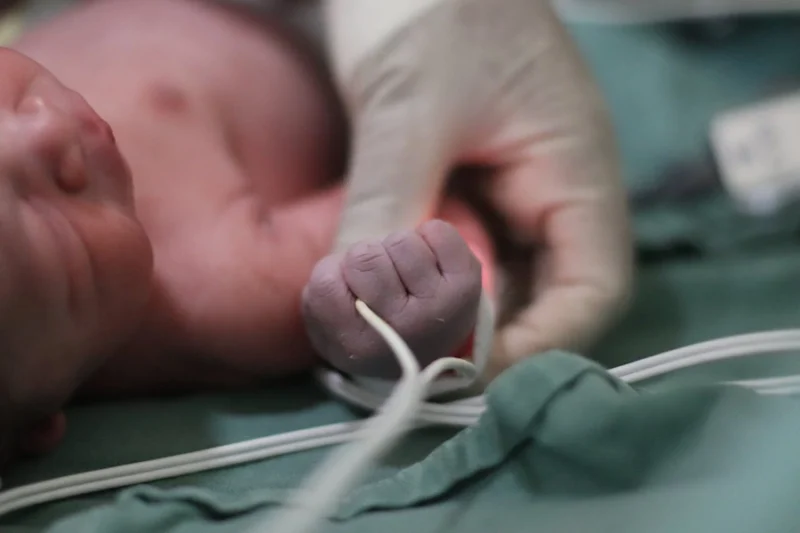
In newborns, signs include low urine output (oliguria), a swollen belly from fluid buildup, or pale skin from waste accumulation. Premature babies might show lethargy, poor feeding, or vomiting. Infections join in, causing fever, cloudy urine with a strong smell, or fussiness.
As children age, recurrent urinary tract infections bring pain during urination, backaches, or nausea. High blood pressure or protein in urine signals advancing issues. In severe forms, like bilateral agenesis, Potter sequence appears: flattened features from low amniotic fluid, breathing troubles, and often early death.
Patients might feel ongoing tiredness, headaches from hypertension, or swelling in legs from fluid retention. Growth delays occur if kidney function drops, affecting bone health and height.
How CAKUT starts and what patients feel
CAKUT begins in the womb, often without maternal awareness. Disruptions hit during key growth windows—early for structural absences, later for obstructions. It starts asymptomatically in most, but pressure from blockages or infections builds discomfort.
Patients with mild forms feel normal until a trigger like dehydration sparks an infection, causing sharp flank pain or burning urination.
In kids, this might show as bedwetting or refusal to eat. For those with cysts or swelling, a dull ache in the side persists, worsening with activity. Neurological effects from toxin buildup include confusion or seizures in extreme cases.
First symptoms of CAKUT
The earliest clues often come prenatally via ultrasound showing enlarged kidneys or low fluid. At birth, failure to urinate within 24 hours, a palpable mass, or visible deformities like in prune-belly syndrome (wrinkled abdomen) alert doctors.
Infections in the first weeks bring fever and irritability.
In undetected cases, a routine check or UTI at 6-12 months reveals the issue.
How CAKUT is diagnosed
Diagnosis starts with imaging. Prenatal ultrasounds spot 80% of cases through hydronephrosis or cysts. Post-birth, kidney ultrasound checks size, position, and structure.
- Voiding cystourethrogram tests for reflux by filling the bladder with contrast.
- Nuclear scans like DMSA assess scarring, while MAG-3 evaluates blockages.
- Lab tests measure creatinine for function, urine for infection or protein, and glomerular filtration rate.
- Genetic testing, via panels or whole-exome sequencing, identifies mutations in complex cases.
- MRI or CT provides detailed views for surgery planning.
Differential diagnosis rules out syndromes like Meckel-Gruber or Bardet-Biedl through exams and genetics.
| Common Diagnostic Tests for CAKUT | Purpose | When Used |
|---|---|---|
| Ultrasound | Views kidney size, cysts, swelling | Prenatal/postnatal screening |
| Voiding Cystourethrogram | Detects urine backflow | After UTI or hydronephrosis |
| DMSA Scan | Checks for scars/damage | In suspected reflux nephropathy |
| Genetic Testing | Identifies mutations/CNVs | Syndromic or familial cases |
| Blood/Urine Labs | Measures function/infection | Routine monitoring |
How CAKUT is treated
Treatment tailors to the anomaly and symptoms. Mild cases need monitoring with ultrasounds and labs to track function. Infections get antibiotics; hypertension, medications like ACE inhibitors (post-infancy).
- Surgery corrects blockages—pyeloplasty for ureteropelvic issues, valve ablation for urethral obstructions, or transplants for failure. Dialysis supports advanced disease.
- Diet limits salt and protein to ease kidney load, especially in kids to support growth.
For prevention, prenatal care avoids risks like diabetes control. Genetic counseling helps families plan.
Multidisciplinary teams—nephrologists, urologists, dietitians—improve long-term results, delaying dialysis and boosting quality of life.
Prognosis varies: mild forms resolve, but severe ones lead to kidney failure in 15-50% by adulthood. Early care extends healthy years.
| Treatment Options by CAKUT Type | Approach | Outcomes |
|---|---|---|
| Vesicoureteral Reflux | Antibiotics, surgery if severe | Many resolve by age 5 |
| Posterior Urethral Valves | Valve removal | 50% progress to CKD in 10 years |
| Renal Agenesis (Unilateral) | Monitoring, protect healthy kidney | Good with compensation |
| Dysplasia/Cysts | Observation, dialysis if failure | Variable, risk of hypertension |
| Obstructions | Surgical repair | Prevents further damage if early |