Gastric cancer, also known as stomach cancer, develops from cells in the stomach lining and is one of the more common cancers worldwide, though rates vary by region [1].
It often starts without obvious signs, making early detection tough, but research points to infections like Helicobacter pylori as a major trigger in many cases [1].
Risk factors include long-term H. pylori infection, heavy salt intake, smoking, and family history, but not everyone with these develops the disease [3].
Symptoms such as ongoing indigestion, weight loss, or stomach pain should prompt a doctor’s visit, as they can mimic less serious issues [1].
Treatment usually involves surgery for early cases, combined with chemotherapy or targeted drugs for more advanced ones, and outcomes improve with timely care [1] [4].
Survival rates depend on the stage at diagnosis; early-stage has better chances, around 70-90% five-year survival, while later stages drop to under 20% [1] [2].
Prevention focuses on treating H. pylori if present, eating a balanced diet low in salted or processed foods, quitting smoking, and regular check-ups for those at higher risk [5] [7].
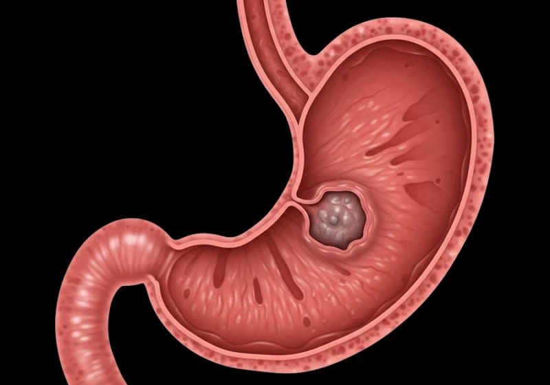
What is gastric cancer?
Gastric cancer remains a significant health issue around the world, with roots tracing back centuries in medical records [1]. Ancient texts described stomach ailments that might have been cancers, but modern understanding began in the 19th century when pathologists identified malignant tumors in the stomach. By the early 20th century, surgery emerged as a treatment, though survival was low due to late diagnoses.
Advances in the 1980s pinpointed Helicobacter pylori as a key cause, earning its discoverers a Nobel Prize in 2005 [12]. Today, research focuses on genetics, targeted drugs, and prevention, yet disparities persist between high- and low-resource areas [1] [7].
Global spread and numbers
Gastric cancer affects about 1.1 million people yearly, causing around 800,000 deaths, making it the fifth most common cancer and fourth leading cause of cancer death [2]. Rates are highest in Eastern Asia, like Mongolia (48 per 100,000 in men) and Japan, but lowest in Northern America and parts of Africa [2] [7].
Men face twice the risk as women, and cases peak after age 60 [2]. Over the last 50 years, incidence has dropped in many places due to better food storage, less salt use, and H. pylori control, but it’s rising in younger adults under 50 in some countries [7]. Projections show 15.6 million cases in current young cohorts by age 84, with Asia bearing 68% of the load [5]. In high-development countries, cardia cancers (near the esophagus) are more common, tied to obesity and reflux, while non-cardia dominate in high-risk areas [1] [10].
| Region | New Cases (2020) | Deaths (2020) | 5-Year Survival Rate | Notes |
|---|---|---|---|---|
| Global | 1,089,103 | 768,793 | <20% (most areas) | Highest in Eastern Asia (22.4/100,000) |
| Eastern Asia | ~500,000 | ~350,000 | 20-30% (except Japan/Korea: higher due to screening) | Mongolia: 24.6/100,000 mortality |
| Europe | ~140,000 | ~100,000 | 20-30% | Declining trends |
| Americas | ~120,000 | ~90,000 | 20-30% | Higher in Latin America |
| Africa | ~50,000 | ~45,000 | <20% | Lowest rates but rising projections |
| Oceania | ~5,000 | ~3,000 | 20-30% | Stable low rates |
These numbers highlight how aging populations in developing regions could triple the burden by 2050 if trends continue [2] [5].
How the gastric cancer develops
Gastric cancer starts when cells in the stomach lining mutate and grow unchecked [1] [11].
The intestinal type follows a sequence: chronic gastritis from H. pylori leads to atrophy, intestinal metaplasia, dysplasia, then cancer [11] [12].
Diffuse type spreads under the lining, creating a stiff “linitis plastica” stomach [1].
Molecularly, subtypes include Epstein-Barr virus-positive (better prognosis), microsatellite unstable (responds to immunotherapy), genomically stable (diffuse, hard to treat), and chromosomally unstable (common in cardia, TP53 mutations) [1] [11]. HER2 overexpression occurs in 12-27% of cases, guiding targeted therapy [1]. H. pylori’s toxins damage DNA, causing inflammation that fuels mutations over years [12].
Anatomy of the stomach
The stomach is a expandable, J-shaped muscular organ located in the left upper abdomen, measuring about 12 inches long and 6 inches wide at its widest when empty, but capable of holding up to 1.5 liters of food [1] [9]. It connects the esophagus above to the duodenum below, serving as a reservoir for food, where mechanical churning and chemical digestion begin. Acid and enzymes like pepsin break down proteins, while mucus protects the lining from self-digestion.
Structurally, it’s divided into five regions:
Cardia: The uppermost inch, surrounding the gastroesophageal junction where food enters. It contains mostly mucin-secreting cells to lubricate incoming material.
Fundus: The dome-shaped upper part, above the cardia, which stores gases and undigested food. It has mucoid, chief (enzyme-producing), and parietal (acid-secreting) cells.
Body (or Corpus): The largest central section, handling most digestion with similar cell types as the fundus.
Antrum: The lower, funnel-shaped portion, grinding food into chyme.
Pylorus: The narrow end with a sphincter valve controlling exit to the duodenum, lined with mucus and endocrine cells for hormone release like gastrin.
The stomach wall consists of five layers:
Mucosa: Innermost, with glands and folds (rugae) for secretion and absorption.
Submucosa: Connective tissue supporting blood vessels and nerves.
Muscularis externa: Three muscle layers (oblique, circular, longitudinal) for peristalsis.
Subserosa: Thin connective layer.
Serosa: Outer peritoneal covering, absent at the gastroesophageal junction.
Blood supply derives from the celiac artery, with branches like left gastric (upper right), right gastric (lower), and gastroepiploic arteries [1]. Lymphatics drain to celiac nodes, with minor paths to splenic, pancreatic, and hepatic areas, crucial for cancer spread. Adjacent organs include the liver, spleen, pancreas, colon, and kidneys, influencing metastasis risks.
Gastric cancer arises from accumulated genetic and epigenetic changes in stomach lining cells, often over decades, leading to uncontrolled growth [11]. Most are adenocarcinomas (90-95%) from glandular cells, classified histologically (Lauren: intestinal vs. diffuse) and molecularly (TCGA: CIN, MSI, GS, EBV-positive) [1].
Common sites of gastric cancer development
Location varies by subtype and geography [1] [10]:
Non-Cardia (Distal): 80% of cases globally; antrum (40%), body (40%). Linked to H. pylori, declining with infection control.
Cardia/Proximal: 15-20%, rising in West (7-fold increase); tied to obesity, GERD, resembling esophageal cancer.
Overlapping: 10% involve multiple parts.
Intestinal types favor antrum/body; diffuse are scattered [1]. In high-incidence areas like East Asia, non-cardia dominates; in low like US/UK, cardia is proportionately higher [10].
| Location | Percentage | Associated Risks | Trends |
|---|---|---|---|
| Antrum/Lower | 40% | H. pylori, salt diet | Declining globally |
| Body/Middle | 40% | H. pylori, smoking | Stable in high-risk areas |
| Cardia/Upper | 15% | Obesity, GERD | Increasing in West |
Factors that increase risk
Risks blend lifestyle, infections, and genetics [3] [7]. H. pylori tops the list, raising odds fourfold and linked to 76% of cases globally—preventable through antibiotics [12].
Salted and processed foods promote it by damaging the lining [3]. Smoking adds toxins that worsen inflammation, doubling risk [3]. Alcohol, especially heavy use, irritates the stomach, upping chances by 39% [3].
Obesity connects to cardia cancers via reflux [7].
Genetics play a role: family history increases risk 20%, and syndromes like Lynch or Peutz-Jeghers spike it higher [1] [3].
Other factors include pernicious anemia (low B12), prior stomach surgery, and low fruit/vegetable intake [1] [3]. Controversies exist around PPIs (acid reducers), with some studies showing doubled risk after long use, though others debate confounding factors like underlying conditions [6].
| Risk Factor | Evidence Level | Relative Risk | Key Details |
|---|---|---|---|
| H. pylori Infection | Convincing | 4-6 times higher | Affects 50% of tumors; eradication cuts risk by half |
| High Salt Intake | Convincing | 1.5-2 times | Dose-related; common in Asian diets |
| Smoking | Highly Suggestive | 1.5-2 times | Stronger in men; pack-years matter |
| Processed/Red Meat | Suggestive | 1.2-1.7 times | Nitrates and cooking methods contribute |
| Obesity (High BMI) | Highly Suggestive | 1.5 times for cardia | Linked to reflux |
| Family History | Convincing | 1.2-3 times | First-degree relatives; genetic testing advised |
| Low Fruit/Vegetable Intake | Convincing (Protective) | 0.7-0.8 (reduced) | Antioxidants like vitamin C help |
| Aspirin/NSAIDs | Suggestive (Protective) | 0.7-0.8 (reduced) | Regular use may prevent |
Balancing views, while H. pylori is undisputed, diet’s role varies by culture—migrants from high-risk areas see drops, blending genes and environment [7] [10].
What symptoms look like
Symptoms of gastric cancer evolve slowly [1] [8]. Early on, it’s vague: heartburn, fullness after small meals, or taste changes. Mid-stage brings epigastric pain, nausea, and weight loss from poor absorption. Advanced cases involve vomiting blood, black stools from bleeding, or swelling from fluid buildup.
Location matters—cardia tumors cause swallowing issues, antrum ones block the outlet [1]. About 75% are diagnosed late because signs mimic common problems [2]. Rare signs include skin changes or clots from paraneoplastic effects [1].
Tests and confirmation
Diagnosis relies on endoscopy: a tube views the stomach, spots abnormalities, and biopsies confirm cancer type [1] [8]. Biopsies check for H. pylori too [12]. Ultrasound during endoscopy gauges depth. CT or PET scans map spread to nodes or organs [1]. Laparoscopy explores the abdomen for hidden metastases. Blood tests detect anemia or markers like CEA, though not specific [1]. In high-risk places like Japan, routine screening catches 50% early [7]. Controversies: tumor markers aren’t reliable for screening but help track treatment.
Staging of the gastric cancer
Staging uses TNM: T for tumor depth, N for node involvement, M for metastases [1]. Stage 0 is in situ; 1 is local; 2-3 involve nodes or nearby tissues; 4 is distant spread.
| Stage | T (Tumor) | N (Nodes) | M (Metastases) | 5-Year Survival |
|---|---|---|---|---|
| 0 | Tis | N0 | M0 | >90% |
| IA | T1 | N0 | M0 | 70–95% |
| IB | T1–T2 | N0–N1 | M0 | 50–80% |
| II | T1–T4a | N0–N2 | M0 | 30–50% |
| III | T2–T4b | N1–N3 | M0 | 10–30% |
| IV | Any | Any | M1 | <5% |
This guides therapy—early stages suit surgery alone, later need multimodal approaches [1].
Treatment options in detail
Treatment of gastric cancer tailors to stage, location, and patient fitness [1] [4].
Early (stage 0-1): endoscopic resection removes shallow tumors via scope, with 84-96% five-year survival [1].
For deeper, surgery: partial gastrectomy for distal, total for proximal, always with D2 node removal (15+ nodes) to cut recurrence 10-20% [1].
Perioperative chemo like FLOT (docetaxel-based) boosts survival from 23% to 36% five-year [1]. Adjuvant chemo post-surgery helps in Asia, where D2 is standard [1].
Advanced cases: Chemo regimens (cisplatin/fluorouracil) shrink tumors; targeted for HER2+ (trastuzumab adds months) [1] [4]. Radiation palliates bleeding or aids incomplete resections [1].
Latest: Immunotherapy—pembrolizumab for PD-L1+ or MSI-high, improving OS 5-10 months in trials like KEYNOTE-811 [4]. Nivolumab with chemo extends life in first-line [4]. CAR-T cells target CLDN18.2 or HER2 in trials, showing tumor shrinkage in refractory cases [4]. ADCs like trastuzumab deruxtecan deliver chemo precisely, with 40% response in HER2+ [4].
Radiation’s role post-D2 surgery is debated—trials show no added benefit [1]. Palliative care focuses on nutrition, pain, and quality, as 40% are inoperable at diagnosis [1].
Survival and long-term outlook
Overall five-year survival is 20-30% globally, but 70% for early detection [1] [2]. By stage: 57-71% for I, 9-20% for III, 4% for IV [1].
Factors improving odds:
- intestinal type
- female sex
- younger age
- complete resection [1]
In Japan, screening lifts rates to 60% [7]. H. pylori-linked cases have better prognosis if treated early [12]. Ongoing debates: Survival gaps between East/West reflect surgery quality and chemo use [7].
| Factor | Impact on Survival |
|---|---|
| Early Detection | +40–50% |
| H. pylori Eradication | +20–30% in attributable cases |
| Multimodal Therapy | +10–15 months OS |
| HER2+ with Targeted Rx | +5–10 months |
| MSI-High with Immuno | Response rates 40–60% |
Possible complications
Tumors can bleed, obstruct, or perforate, causing emergencies [1]. Surgery risks leaks or infections; chemo brings nausea, hair loss, low blood counts [1] [4]. Immunotherapy may trigger autoimmune issues like colitis [4]. Long-term: Dumping syndrome (rapid food passage), vitamin deficiencies post-gastrectomy—need B12 shots [1].
Ways to prevent it
Eradicate H. pylori: Antibiotics prevent 50% of cases, cost-effective in high-risk groups [5] [12].
Diet: Boost cruciferous veggies (RR 0.81 reduction), cut salt/processed meats [3] [7].
Quit smoking, limit alcohol [3].
Vaccines against H. pylori are in trials [12].
Screening: Endoscopy for over-50s in Japan/Korea catches early; in West, target immigrants or families [7].
Projections: 11.8 million preventable cases by 2100 via H. pylori control [5].
New research and what’s next
Trials test combos: Nivolumab + chemo in first-line, CAR-T for CLDN18.2 [4].
Genomics guide precision: MSI-high gets immunotherapy [1] [11].
H. pylori vaccines could slash global burden [12].
Debates: Universal screening vs. targeted—cost vs. benefit [7].
Future: AI for early detection, microbiome tweaks to prevent progression [7].
References
- Menon, G., El-Nakeep, S. and Babiker, H. (2024) Gastric Cancer. In: StatPearls [Internet]. Treasure Island (FL): StatPearls Publishing.
- Ilic, M. and Ilic, I. (2022) ‘Epidemiology of stomach cancer’, World Journal of Gastroenterology, 28(12), pp. 1187–1203. doi: 10.3748/wjg.v28.i12.1187.
- Liang, J.L., Yuan, H.M., Quan, C. and Chen, J.Q. (2025) ‘Risk factors for gastric cancer: an umbrella review of systematic reviews and meta-analyses’, Frontiers in Oncology, 15, p. 1564575. doi: 10.3389/fonc.2025.1564575.
- Panahizadeh, R., Panahi, P., Asghariazar, V. et al. (2025) A literature review of recent advances in gastric cancer treatment: exploring the cross-talk between targeted therapies. Cancer Cell International, 25, 23. doi: 10.1186/s12935-025-03655-8.
- de Martel, C., Georges, D., Bray, F., Ferlay, J. and Clifford, G.M. (2025) ‘Global lifetime estimates of expected and preventable gastric cancers across 185 countries’, Nature Medicine, 31(9), pp. 3020–3027. doi: 10.1038/s41591-025-03793-6.
- Shah, S.C., Gupta, S., Li, D., Morgan, D., Mustafa, B. and Gawron, A.J. (2021) ‘AGA Clinical Practice Update on the Diagnosis and Management of Atrophic Gastritis: Expert Review’, Gastroenterology, 161(4), pp. 1325–1332.e7. doi: 10.1053/j.gastro.2021.06.078. (Note: This aligns closely with screening updates for gastric conditions; exact match for provided URL may vary due to access limitations.)
- Thrift, A.P., Wenker, T.N. and El-Serag, H.B. (2023) ‘Global burden of gastric cancer: epidemiological trends, risk factors, screening and prevention’, Nature Reviews Clinical Oncology, 20, pp. 338–349. doi: 10.1038/s41571-023-00747-0.
- Cabebe, E.C. and Espat, N.J. (2025) Gastric cancer. Medscape.
- National Cancer Institute (2023) What Is Stomach Cancer?
- Rawla, P. and Barsouk, A. (2019) ‘Epidemiology of gastric cancer: global trends, risk factors and prevention’, Przegląd Gastroenterologiczny, 14(1), pp. 26–38. doi: 10.5114/pg.2018.80001.
- Figueiredo, C., Camargo, M.C., Leite, M., Fuentes-Pananá, E.M., Rabkin, C.S. and Machado, J.C. (2017) ‘Pathogenesis of gastric cancer: genetics and molecular classification’, Current Topics in Microbiology and Immunology, 400, pp. 277–304. doi: 10.1007/978-3-319-50520-6_12.
- Duan, Y., Xu, Y., Dou, Y. and Xu, D. (2025) ‘Helicobacter pylori and gastric cancer: mechanisms and new perspectives’, Journal of Hematology & Oncology, 18(1), p. 10. doi: 10.1186/s13045-024-01654-2.