Allodynia is the occurrence of pain sensations in response to stimuli that normally do not cause pain. It is noted in causalgia, neuropathies and polyneuropathies, herpes zoster, multiple sclerosis, migraine, conditions after spinal cord injuries, strokes, and spinal surgeries. To determine the cause of allodynia, data from interviews, general and neurological examinations, electrophysiological, neuroimaging, and laboratory tests are used. Treatment includes analgesics, blocks with anesthetics, vitamins, antidepressants, physiotherapeutic methods. Sometimes operations are recommended.
General information

Allodynia is pain arising from the action of non-painful stimuli. It differs from hyperalgesia, where the pain response occurs in response to an appropriate stimulus but is excessive. It can be provoked by touch, wind, non-intense thermal effects, and so on. It is one of the variants of neuropathic pain, observed in 15-50% of people with this syndrome.
In 83-94% of cases, it is due to peripheral nerve damage, and in 6-17%, it is a consequence of pathological processes in the central nervous system (CNS). There are such types of allodynia as:
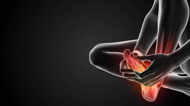
Mechanical allodynia
The most common. Noted with tactile stimuli. Can be static or dynamic. The first variant is observed with single touches (for example, an attempt to take someone by the hand), the second – with repeated weak stimulation (for example, repeated movements of a brush or cotton pad on the skin when applying or removing makeup).
Thermal allodynia
Develops under the influence of cold and hot stimuli that normally do not cause discomfort. Allodynia can be observed when taking a bath or shower, touching warm or cool objects, going outside from indoors.
Muscular
Unlike the two previous variants, it is associated not with skin stimulation but with movements. Patients complain of pain deep in the soft tissues (in the projection of muscles, joints, ligaments, tendons) in the absence of organic changes in these anatomical structures.
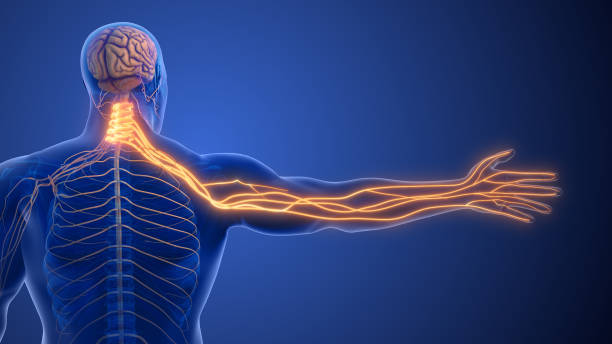
Pathophysiological mechanisms
Allodynia results from complex alterations in pain processing pathways occurring at multiple levels of the nervous system. Understanding these mechanisms helps explain why non-painful stimuli can trigger pain sensations and guides the development of targeted therapeutic approaches.
Peripheral sensitization
In peripheral sensitization, damaged or inflamed peripheral nerves become hyperexcitable due to:
- Ion Channel Alterations: Injury to peripheral neurons leads to upregulation and redistribution of voltage-gated sodium channels (particularly Nav1.7, Nav1.8, and Nav1.9), resulting in decreased activation thresholds and increased membrane excitability. This causes neurons to fire in response to normally subthreshold stimuli.
- Inflammatory Mediator Effects: Following tissue damage, inflammatory mediators including prostaglandins, bradykinin, serotonin, and various cytokines (IL-1β, IL-6, TNF-α) bind to receptors on nociceptors, triggering intracellular signaling cascades that phosphorylate receptor proteins and ion channels, lowering their activation thresholds.
- Phenotypic Switching: Low-threshold Aβ mechanoreceptors, which normally respond to light touch and vibration, begin expressing neuropeptides typically found in nociceptive C-fibers. This causes mechanoreceptors to function more like pain fibers, contributing to touch-evoked pain.
- Sprouting of Nerve Fibers: Following nerve injury, aberrant sprouting and reorganization of nerve fibers can create inappropriate connections between tactile and pain pathways.
Central sensitization
Central sensitization involves neuroplastic changes in the central nervous system, particularly at the spinal cord and brain level:
- Wind-up Phenomenon: Repeated activation of C-fibers leads to progressively increasing responses in dorsal horn neurons due to temporal summation of slow synaptic potentials, primarily mediated by NMDA receptors.
- Glutamate Receptor Modulation: Persistent nociceptive input causes upregulation and enhanced phosphorylation of NMDA and AMPA receptors, increasing neuronal excitability in pain processing pathways.
- Loss of Inhibitory Control: Reduced activity of inhibitory interneurons containing GABA and glycine in the dorsal horn removes the natural “brakes” on pain transmission. This disinhibition can result from death of inhibitory interneurons or reduced effectiveness of descending pain modulatory pathways.
- Glial Cell Activation: Microglia and astrocytes become activated following nerve injury and release pro-inflammatory cytokines, chemokines, and growth factors that enhance neuronal excitability and contribute to central sensitization.
- Synaptic Remodeling: Long-term potentiation (LTP) at synapses in pain pathways strengthens connections between neurons, similar to memory formation. This creates a “pain memory” that can persist even after the original injury has healed.
Neuronal circuit reorganization
In chronic conditions leading to allodynia, substantial reorganization occurs in sensory processing circuits:
- Spinal Cord Reorganization: Low-threshold Aβ fibers that normally terminate in deeper laminae of the dorsal horn sprout into superficial laminae (I-II) where nociceptive information is processed. This anatomical reorganization allows touch information to directly access pain-processing circuits.
- Cortical Reorganization: Chronic pain conditions cause functional and structural changes in somatosensory cortex, thalamus, and limbic structures, altering the brain’s interpretation of sensory stimuli.
- Altered Descending Modulation: Dysfunction in descending pain modulatory pathways from the brainstem (including periaqueductal gray, rostral ventromedial medulla, and locus coeruleus) reduces the ability to suppress inappropriate pain signals.
Neurotransmitter and neuromodulator imbalance
Various neurotransmitters and neuromodulators play crucial roles in allodynia development:
- BDNF Signaling: Brain-derived neurotrophic factor released from microglia causes disinhibition in the spinal dorsal horn by disrupting chloride homeostasis in neurons.
- Substance P and CGRP: These neuropeptides increase in concentration and facilitate pain transmission through enhanced release from primary afferents and increased receptor expression in the dorsal horn.
- Serotonin and Norepinephrine: Imbalances in these neurotransmitters in descending modulatory pathways contribute to insufficient pain inhibition from higher centers.
Physiological causes of allodynia
In healthy people, allodynia is provoked by excessive irritation of a particular area. Most often, the head is affected. In women, the symptom is a consequence of using a hairdryer, abusing thermal styling, sleeping with hair curled on hard curlers. The cause of unpleasant sensations in people of both sexes is prolonged wearing of tight headwear, sometimes – pediculosis (primarily in children).
Temporary allodynia in the distal parts of the lower extremities is potentiated by tight or uncomfortable shoes. In summer, pain with non-painful stimuli develops against the background of mild sunburn. In people who have undergone injuries and surgical interventions, allodynia in the area of the scar and adjacent tissues is often observed in the period after suture removal, disappearing as the final healing and restoration of small nerves and nerve endings occur.
Fibromyalgia
More common in middle-aged women, it develops against a background of hereditary predisposition, accompanied by constant diffuse pain in the body, often in combination with numbness, tingling, and other sensory phenomena, predominantly expressed in the limbs. Allodynia and spontaneous pain sensations cause fatigue, sleep disturbances, and depression. Provoking factors for fibromyalgia are:
- Conditions after injuries: burns, bruises, fractures, damage to peripheral nerve trunks.
- Infectious diseases: Q fever, infectious mononucleosis, borreliosis.
- Endocrine disorders: hypothyroidism.
- Distress: psychoemotional exhaustion due to acute and chronic stressful situations.
- Medical interventions: taking certain medications, vaccination.
Polyneuropathies
The clinical picture of polyneuropathy consists of sensory, motor (paresis), and autonomic (dry skin, vascular regulation disorders) symptoms. Allodynia and other sensory disorders (paresthesias, hyperesthesia) are due to damage to thin myelinated fibers, occur at the initial stage, and are later replaced by hypoesthesia.
Most cases of allodynia are associated with the development of peripheral diabetic polyneuropathy. The pathology is found in more than 10% of patients with diabetes mellitus. The symptom is also determined in the following polyneuropathies:
- Toxic: alcoholic, in drug addictions, poisonings, occupational hazards.
- Infectious: in measles, mumps, diphtheria, HIV infection.
- Autoimmune: paraneoplastic, paraproteinemic, in Miller-Fisher syndrome.
- Metabolic: in renal and hepatic insufficiency.
- Hereditary: in Roussy-Levy syndrome, Refsum’s disease, and Charcot-Marie-Tooth disease.
- Others: drug-induced, arising after chemotherapy.
Neuralgias
The most common pathology in this group is trigeminal neuralgia. There are multiple attacks of extremely intense prosopalgia, which spreads to half of the face and resembles an electric shock. When shaving, contacting cold water, going outside, exposure to wind, allodynia develops, turning into another paroxysm. Other possible causes of the symptom are the following neuralgias:
- Auricular ganglion. Pulsating or burning pain occurs in the ear and parotid area. It can be potentiated by hypothermia of the affected area, eating hot food, experiences, emotional tension.
- Submandibular and sublingual ganglia. Allodynia develops when eating cold or hot food. It transforms into a pain attack in the submandibular and sublingual areas, anterior parts of the tongue.
- Intercostal. Paroxysms are caused by movements of the chest (coughing, laughing, deep breaths, sharp turns of the torso), less often – by tactile contacts. The pain has a girdling character, spreading along the intercostal space.
Causalgia
Occurs after traumatic injuries (fractures, gunshot wounds). At the initial stage, causalgia manifests as burning or stinging pain that does not correspond to the severity of the injury. Painful sensations are provoked by any stimuli (movement, touch), reduced by immersing the affected segment in water or applying a wet bandage. Allodynia is accompanied by hyperpathia, hyperalgesia, edema, trophic disorders. Over time, paresis and contractures form.
Herpes Zoster
Herpes zoster manifests with nonspecific prodromal signs. Then itching and pain appear along the course of the peripheral nerve (in the vast majority of cases – intercostal). After a few days, painful pink spots form in the affected area, and a little later – vesicles with serous content. After recovery, approximately 10% of patients retain allodynia and other manifestations of neuropathic pain for several years.
Cephalalgias
Periodic skin allodynia in the temple area is observed in 65% of patients with migraine. In 20% of cases, it causes significant discomfort, has an additional negative impact on quality of life. Patients complain of periodic cephalalgias that cover half of the head, accompanied by nausea, fear of bright light and loud sounds. Allodynia is also found in paroxysmal hemicrania, combined with pain and autonomic disorders.
Other causes
Allodynia, due to organic changes in the central nervous system, can be observed in such pathological processes as:
- Syringomyelia;
- Post-stroke condition;
- Multiple sclerosis (with the occurrence of acute transverse myelitis);
- Condition after spinal cord injuries and surgical interventions on the spine.
In addition, allodynia is found in combined pain syndromes developing against the background of impaired nociceptive mechanisms with the participation of psychogenic and neuropathic components. A common example is chronic pain in radiculopathy due to degenerative and inflammatory diseases of the spine. Less commonly, allodynia occurs with the ingrowth of nerves by malignant tumors.
Diagnosis
The cause of allodynia is determined by a neurologist. In the development of neuropathic pain against the background of other pathologies, consultations with an endocrinologist, therapist, narcologist, and so on may be indicated. As part of the interview, the doctor finds out the time of appearance of the symptom, asks about other manifestations, tracks the dynamics of the disease development. Collects life history to identify possible provoking factors. The additional examination program includes the following procedures:
Neurological examination
The neurologist determines the localization and type of allodynia using special tests (by ordinary touch, using a brush, warm and cold objects). Assesses various types of sensitivity, identifies sensory disorders. Examines muscle strength and reflexes.
Electrophysiological methods
EMG and ENG are performed to assess the functional state of the nerve, determine the acuity of the process, the level and degree of damage to nerve trunks, and monitor recovery during treatment. Recommended for neuralgias, polyneuropathies, allodynia of central origin.
Neuroimaging
For neuralgias, MRI of the brain and CT of the skull may be prescribed to clarify the cause of nerve damage (compression in a narrow bone canal, tumor). If vascular compression is suspected, MR-angiography is performed. In conditions after injuries and operations on the spine, MRI of the spinal cord and CT of the vertebral column are indicated. Neuroimaging methods are also used to diagnose CNS diseases accompanied by allodynia.
Laboratory tests
In fibromyalgia, a decrease in the level of serotonin and L-tryptophan in the blood is determined. In metabolic polyneuropathies, hyperglycemia, signs of liver or kidney dysfunction are found according to blood biochemistry, in autoimmune – corresponding antibodies. In the infectious etiology of allodynia, PCR, ELISA, microbiological examination are required.
Treatment
Conservative therapy
The treatment tactics are determined taking into account the etiology of allodynia:
- Fibromyalgia.
For acute pain, central analgesics are used. To reduce the severity of chronic pain syndrome, anticonvulsants are prescribed, to eliminate depression and improve sleep – antidepressants. It is possible to introduce local anesthetics into trigger points, irrigation of pain areas. As part of non-drug treatment, psychotherapy, exercise therapy, hydrotherapy, acupuncture, biofeedback therapy are recommended.
- Polyneuropathies.
For intense pain, tricyclic antidepressants are indicated. In diabetes, the insulin therapy regimen is adjusted, in uremia, hemodialysis is performed, in intoxications, detoxification therapy is carried out, in inflammatory polyneuropathies, membrane plasmapheresis is performed, human immunoglobulin is administered. Symptomatic treatment includes B vitamins, neurotrophic agents.
- Trigeminal Neuralgia.
To eliminate hyperexcitation, anticonvulsants are used. To increase the effectiveness of the main drug, medications with antihistamine action and microcirculation correctors are used. Pain paroxysms are stopped with spasmolytics. Therapeutic blocks with local anesthetics and glucocorticoids are performed. Analgesic and anti-inflammatory effects are provided by physiotherapeutic procedures: diadynamic currents, ultraphonophoresis with hydrocortisone, galvanization with novocaine.
- Other Neuralgias.
The list of used medications includes adrenergic blockers, ganglionic blockers, blockers of cholinoreactive systems, ATP, vitamins C, PP, B. To normalize the psychoemotional state and reduce pain, antidepressants, tranquilizers, sedatives are indicated. During the convalescence period, biogenic agents, reflexotherapy, inductothermy, DDT, galvanization, SMT, ultraphonophoresis are recommended.
- Migraine.
For paroxysms of moderate and mild intensity, combined and simple analgesics, codeine-containing agents are prescribed. Sometimes therapeutic blocks are performed. Severe attacks are an indication for oral treatment or subcutaneous administration of triptans.
Surgical treatment
In case of damage to the sublingual or submandibular ganglia, ENT operations (frontotomy, maxillary sinusotomy, tonsillectomy) may be required to eliminate the focus of infection. Patients with involvement of the auricular ganglion are sometimes indicated for sanitizing surgery on the middle ear. In trigeminal neuralgia, microsurgical decompression or percutaneous radiofrequency destruction is performed. In some cases, the sensory root is destroyed using stereotactic surgery methods.
Allodynia Prognosis by Condition
| Condition | Prognosis | Recovery Timeline | Long-term Outlook |
|---|---|---|---|
| Physiological Transient Allodynia | Excellent | Resolves within 24-72 hours after removing stimulus | Complete resolution; low risk of recurrence if precipitating factors are avoided |
| Post-traumatic and Post-surgical | Generally good | Significant improvement within 3-6 months; residual symptoms may persist in up to 20% for 12-18 months | Complete resolution in 75-80% of cases within 2 years |
| Herpes Zoster (Shingles) | Age-dependent | Under 50 years: 2-3 months Over 70 years: 30-50% persist beyond one year |
5% develop persistent allodynia lasting more than 5 years; early treatment reduces risk of persistence by 30-40% |
| Diabetic Polyneuropathy | Generally progressive without proper glycemic control | Complete resolution uncommon; management rather than cure is the goal | With optimal management, 50% achieve significant symptom reduction within 6-12 months, though ongoing therapy required |
| Trigeminal Neuralgia | Variable based on treatment | Medical treatment: 70-80% initial response; efficacy decreases to 50% after 5-10 years Surgical interventions: 70-90% initial pain relief |
Characterized by remissions and exacerbations; 10-15% experience natural remission without intervention |
| Multiple Sclerosis-related | Tied to disease course and activity | During acute episodes: improvement over 6-12 weeks with treatment | 60-70% experience recurrence; disease-modifying therapies reduce frequency by 30-50% |
| Fibromyalgia | Chronic condition with fluctuating severity | Symptom reduction begins within 4-8 weeks of treatment initiation | 20-25% achieve long periods of minimal symptoms; 10-15% experience progressive worsening; 60-70% maintain normal activities with management |
| Post-stroke | Often becomes chronic | May develop immediately or up to 6 months post-stroke; 15-20% show spontaneous improvement within first year | 70-80% develop chronic condition requiring ongoing management; early rehabilitation reduces severity in 30% of cases |
Predictors of poor prognosis
Several factors are associated with more persistent allodynia regardless of etiology:
- Severe initial pain intensity
- Delay in diagnosis and appropriate treatment
- Concurrent depression or anxiety disorders
- Poor sleep quality
- Catastrophizing pain coping style
- Advanced age
- Multiple comorbidities
- Female gender (in some conditions)
Predictors of favorable outcomes
Factors associated with better recovery include:
- Early and aggressive multimodal treatment
- Good compliance with therapy
- Active participation in physical rehabilitation
- Implementation of appropriate self-management strategies
- Strong social support systems
- Absence of mood disorders
- Younger age
- Limited duration of symptoms before treatment initiation
Understanding the typical course and prognostic factors helps clinicians provide realistic expectations to patients and develop appropriate long-term management strategies for allodynia based on its underlying cause.
References and sources used to write this article:
- Merskey H, Bogduk N. (2012). Classification of Chronic Pain: Descriptions of Chronic Pain Syndromes and Definitions of Pain Terms (2nd ed.). IASP Press.
- Fields HL, Basbaum AI, Heinricher MM. (2023). Central Nervous System Mechanisms of Pain Modulation. In: McMahon SB, Koltzenburg M, Tracey I, Turk DC, editors. Wall & Melzack’s Textbook of Pain (7th ed.). Elsevier.
- Woolf CJ, Mannion RJ. (2021). Neuropathic Pain: Etiology, Pathophysiology, Mechanisms, and Assessments. In: Bonica’s Management of Pain (5th ed.). Lippincott Williams & Wilkins.
- Jensen TS, Finnerup NB. (2024). Allodynia and hyperalgesia in neuropathic pain: Clinical manifestations and mechanisms. The Lancet Neurology, 23(2), 114-127.
- Basbaum AI, Bautista DM, Scherrer G, Julius D. (2021). Cellular and molecular mechanisms of pain. Cell, 187(3), 267-284.
- Latremoliere A, Woolf CJ. (2020). Central sensitization: A generator of pain hypersensitivity by central neural plasticity. Journal of Pain, 21(10), 895-926.
- Lipton RB, Bigal ME, Ashina S, et al. (2023). Cutaneous allodynia in the migraine population. Annals of Neurology, 93(1), 18-24.
- Oaklander AL, Wilson PR, Moskovitz PA, et al. (2023). Response to first-line treatment as a predictor of persistent post-herpetic neuralgia: A prospective study. Pain Medicine, 24(4), 412-423.
- Truini A, Barbanti P, Pozzilli C, Cruccu G. (2022). A mechanism-based classification of pain in multiple sclerosis. Journal of Neurology, 269(8), 775-782.
- Freeman R, Baron R, Bouhassira D, et al. (2024). Sensory symptoms and signs in diabetes: Clinical and diagnostic aspects. Diabetes Care, 47(4), 802-815.
- Colloca L, Ludman T, Bouhassira D, et al. (2022). Neuropathic pain: Nature Reviews Disease Primers, 8(1), 1-24.
- Finnerup NB, Attal N, Haroutounian S, et al. (2024). Pharmacotherapy for neuropathic pain in adults: A systematic review and meta-analysis. The Lancet Neurology, 23(3), 228-240.
- Dworkin RH, O’Connor AB, Kent J, et al. (2023). Interventional management of neuropathic pain: NeuPSIG recommendations. Pain, 164(11), 2058-2074.
- Haanpää M, Attal N, Backonja M, et al. (2022). NeuPSIG guidelines on neuropathic pain assessment. Pain, 163(5), 1031-1048.
- Sullivan MJL, Thorn B, Rodgers W, Ward LC. (2023). Path model of psychological antecedents to pain experience: Experimental and clinical findings. Clinical Journal of Pain, 39(4), 350-365.
- Price TJ, Cervero F, Gold MS, et al. (2022). Transition to chronic pain: Opportunities for novel therapeutics. Nature Reviews Neuroscience, 23(5), 319-331.
- Tracey I, Woolf CJ, Andrews NA. (2021). Composite Pain Biomarker Signatures for Objective Assessment and Effective Treatment. Neuron, 109(1), 46-64.
- Sandkühler J. (2023). Models and mechanisms of hyperalgesia and allodynia. Physiological Reviews, 103(2), 707-760.
- Choucair-Jaafar N, Beetz C, Gilsbach R, et al. (2023). Voltage-gated sodium channel conformational dynamics as therapeutic targets for pain. Nature Reviews Drug Discovery, 22(1), 57-76.
- Denk F, McMahon SB, Tracey I. (2022). Pain vulnerability: A neurobiological perspective. Nature Neuroscience, 25(2), 246-259.